
Skeletal Muscle Definition
Skeletal muscle is a specialized contractile tissue that moves the body of an organism. The skeletal muscle is made up of bundles of muscle fibers surrounded by protective membranes. As a result of this arrangement, skeletal muscle can contract and release rapidly without being subjected to too much friction. In most multicellular organisms, skeletal muscle tissue exists.
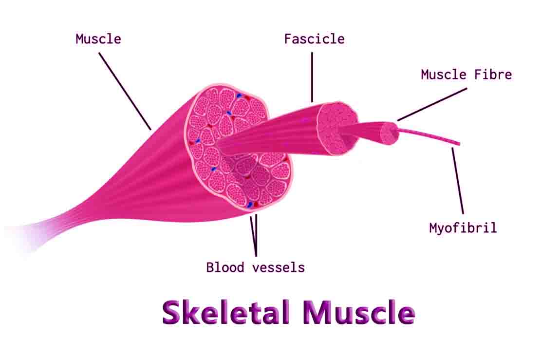
Skeletal Muscle Structure
Muscle fibers made of muscle cells make up skeletal muscle. Muscle cells have multiple nuclei and are long. The end of each skeletal muscle is connected to the bone by a tendon. Skeletal muscle is covered with collagenous epimysium, which connects directly with this tendon. Fascicules are bundles of muscle fibers that lie underneath the epimysium.
Collagen forms another protective covering around these fascicles. Through the perimysium, nerves and blood vessels can pass through the muscle.
There are tens to hundreds of muscle fibers bundled together in each fascicle. Muscle fibers are formed by chains of multinucleated muscle cells. As these fibers are bundled into fascicles, another layer called an endomysium protects them. Under a microscope, each muscle cell has distinct regions. The bands or striae of skeletal muscle are made up of these sarcomeres. An individual sarcomere consists of a complex of proteins that contract the muscle.
Sarcomeres are composed of actin and myosin, as well as helper proteins. Actin and myosin filaments can be seen between the dark bands. In the image above, actin appears as a twisting filament made up of many actin units. Actin is stabilized and provides a pathway for muscle contraction with the help of proteins. In terms of importance, troponin and tropomyosin are the most important.
Actin filaments are surrounded by tropomyosin, which prevents myosin heads from attaching. Until tropomyosin receives the signal to contract, troponin locks it in place. There are many interlaced tails of myosin units in a myosin fiber. Actin filaments attract the heads of the units, which stick above the fiber.
Function of Skeletal Muscle
Your brain sends a nervous signal to your nerves when you want to move your arm. Raising your arm requires many muscles, so the signal is sent down many nerves to many muscles. At neuromuscular junctions, nerve impulses are received by each skeletal muscle. Muscle cells can be stimulated by nerve impulses in these places.
In the sarcolemma, the plasma membrane of skeletal muscle cells, the impulse travels down channels. Channels lead inside cells at certain points in the membrane. Cells contain transverse tubules that carry nervous impulses. An endoplasmic reticulum specialized for calcium ions, the sarcoplasmic reticulum, releases calcium ions. As a result of calcium ions, troponin is released from tropomyosin. A shift in tropomyosin position allows myosin heads to attach to actin filaments.
ATP will be used to contract the filament once the myosin heads are attached. The myosin heads crawl down the filament slowly. The ATP energy is used to move one head while the other is attached. A sarcomere can be contracted up to 70% of its original length when hundreds or thousands of heads are involved.
An arm can lift fluidly as the nervous impulse hits each muscle fiber and muscle at the same time. Every skeletal muscle has special sensory cells that send feedback to the brain as an added feedback measure. A specialized protein in these cells, called muscle spindles, detects tension. As soon as a cell receives tension, it starts a nervous impulse and sends the signal through neurons to the brain.
As a result of piecing together this complex framework of inputs and outputs, the brain is able to determine where the body is in space. We are able to move our bodies in a coordinated manner thanks to the somatic nervous system. Skeletal muscle is controlled almost exclusively by the somatic nervous system, whereas cardiac and smooth muscles are controlled by the autonomous nervous system. It is easy to demonstrate this system. Make several claps with your hands as you close your eyes.
Have your hands met? This is because your brain has been training in coordination since birth, and recognizes the specific tensions on each muscle as you swing your hands. As you clap, these inputs are monitored and adjustments are made to ensure your hands continue to make contact with each other. The same system is responsible for balance, coordination, and most physical movements.
Skeletal Muscle Location
Skeletal muscle, as the name implies, is any muscles that connects to and controls the motions of the skeleton. In all there are somewhere between 600 and 900 muscles in the human body, but an exact number is hard. Many muscles are obscurely small or are sometimes grouped together with similar muscles. Skeletal muscle is found between bones, and uses tendons to connect the epimysium to the periosteum, or outer covering, of bone.
Skeletal muscle is adapted and shaped in many different ways, which give rise to complex movements. Skeletons are not always internal as they are in humans. Even animals with exoskeletons, like crabs and mussels, have skeletal muscle. While the muscle might be adapted differently depending on the animal, skeletal muscle is defined by its striations and connections to skeleton. Everything from the flapping of a bird’s wings to the crawling of a beetle are carried out by skeletal muscle.
FAQ’s
Skeletal muscle is a type of muscle tissue that is attached to bones and helps facilitate voluntary movement of the body.
Skeletal muscle works by contracting and relaxing in response to nerve signals sent by the brain. When a muscle contracts, it pulls on the bones it is attached to, causing movement.
Skeletal muscle is composed of muscle fibers, connective tissue, blood vessels, and nerves.
Connective tissue provides support and structure for skeletal muscle fibers, and helps transmit the force generated by muscle contraction to the bones.
There are two main types of skeletal muscle fibers: slow-twitch (Type I) and fast-twitch (Type II). Slow-twitch fibers are used for endurance activities such as long-distance running, while fast-twitch fibers are used for explosive activities such as sprinting or weightlifting.